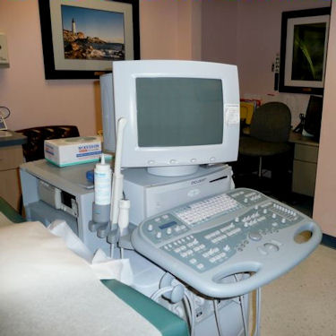
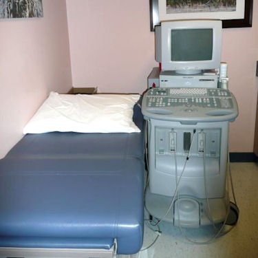
Echocardiogram

Frequently Asked Questions Regarding Ultrasound

Echocardiogram (Echo, 2D Echo, cardiac ultrasound, echocardiography)

The echocardiogram is an ultrasound of the heart. Using standard ultrasound techniques, two-dimensional slices of the heart can be imaged. The M-mode and 2-D Echo evaluates the size, thickness, and movement of the heart structure (chambers, valves, etc.). In addition to creating two-dimensional pictures of the cardiovascular system, the echocardiogram can also produce accurate assessment of the velocity of blood and cardiac tissue at any arbitrary point using pulsed or continuous wave Doppler ultrasound.

Doppler is a special part of the ultrasound examination that assess blood flow (direction and velocity). During the Doppler examination, the ultrasound beam will evaluate the flow of blood as it makes it way though and out of the heart. This information is presented visually on the monitor(as color images or grayscale tracings and also as a series of audible signals with a swishing or pulsating sound). This allows assessment of cardiac valves areas and function, any abnormal communications between the left and right side of the heart, any leaking of blood through the valves (vascular regurgitation), and calculation of the cardiac output as well as the ejection fraction.

Your doctor may perform an echocardiogram to:

  • Assess the overall function of your heart.
  • Determine the presence of may types of heart disease.
  • Follow the progress of heart valve disease over time.
  • Evaluate the effectiveness of medical or surgical treatments.

Patient who are experiencing the following symptoms may need to have a echocardiogram: Chest Pain Shortness of Breath Heart Murmur Hypertension Edema Other symptoms are determined under the doctors discretion.

  • You can wear whatever you like. You will need to change into a gown to wear during the echocardiography.
  • You may eat and drink as you normally would on the day of the test.
  • Take all of your medications at the usual times, as prescribed by your doctor.
  • A cardiac sonographer will place three electrodes (small, flat, sticky patches) on your chest. The electrodes are attached to an electrocardiograph monitor (ECG) that charts your heart’s electrical activity.
  • The sonographer will ask you to lie on your left side on an exam table. The sonographer will place a wand (called a sound-wave transducer) on several areas of your chest. The wand will have a small amount of cool gel on the end, which will not harm your skin. This gel helps get clearer pictures.
  • Sounds are part of the Doppler signal. You may or may not hear the sounds during the test.
    you may be asked to change positions during the exam in order to take pictures of different areas of your heart. You may be asked to hold your breath at times.
  • You should feel no major discomfort during the test. You may feel coolness from the gel on the transducer and a slight pressure of the transducer on your chest.

A brief examination in an uncomplicated case may be done within 15 to 20 minutes. The additional use of Doppler may add an additional 10 to 20 minutes. However, it may take up to an hour when there are multiple problems or when there are technical problems (for example, patients with lung disease, obesity, restlessness, and significant shortness of breath may be more difficult to image).

Echocardiography is an invaluable tool in providing the doctor with important information about the following:

Size of the chambers of the heart, including the dimension or volume of the cavity and the thickness of the walls. The appearance of the walls may also help identify certain types of heart disease that predominantly involve the heart muscle.

Pumping function of the heart can be assessed by echocardiography. One can tell if the pumping power of the heart is normal or reduced to a mild or severe degree. This is known as an ejection fraction or EF. A normal EF is around 55 to 65%. Numbers below 45% usually represent some decrease in the pumping strength of the heart, while numbers below 30 to 35% are representative of an important decrease.

Valve Function: Echocardiography identifies the structure, thickness and movement of each heart valve. It can help determine if the valve is normal, scarred from an infection or rheumatic fever, thickened, calcified (loaded with calcium), torn, etc. It can also assess the function of prosthetic or artificial heart valves. The additional use of Doppler helps to identify abnormal leakage across heart valves and determine their severity. Doppler is also very useful in diagnosing the presence and severity of valve stenosis or narrowing.

Volume status: Low blood pressure can occur in the setting of poor heart function but may also be seen when patient’s have a reduced volume of circulating blood (as seen with dehydration, blood loss, use of diuretics or ‘water pill.”, etc.)

Other Uses: Echocardiography is useful in the diagnosis of fluid in the pericardium (the sac that surrounds the heart). It also determines when the problem is severe and potentially life-threatening. Other diagnoses made by Doppler or echocardiography include congenital heart diseases, blood clots or tumors within the heart, active infection of the heart valves, abnormal elevation of pressure within the lungs, etc.

.

Yes, your doctor must provide a referral (prescription) in order for you to receive a examination. In addition, some insurance carriers or HMO’s require a pre-certification. Please discuss this with your doctor and your insurance company or HMO prior to your test.

If a doctor is present during the test or reviews it while you are still in the office, you may be able to get the results before you leave. However, the doctor is not routinely present during the test and you may have to wait from one to several days before the images have been reviewed by the Cardiologist and the report is generated and sent to the ordering physician by mail. A detailed written report of the procedure, findings, and results will be sent to the ordering physician. Your physician will then call you to discuss the results or you will be instructed to follow up with the ordering physician in the office to review the results. Urgent results will be telephoned immediately to your doctor.

Echocardiography is extremely safe. There are no known risks from the clinical use of ultrasound during this type of testing.